

1. Which of the following statements about Intra-
Cranial Pressure (ICP) monitoring is TRUE?
A. The transducer of the intra-parenchymal pressure
monitor is placed at the level of the tragus
B. The intra-ventricular pressure monitor does not
provide a global measurement of ICP
C. ICP- guided therapy improves outcomes in
patients with traumatic brain injury
D. ICP monitoring is indicated in brain injured
patients with GCS ≤8 even if the CT scan is normal
Next Question

1A. The transducer of the intra-parenchymal
pressure monitor is placed at the level of the tragus
Intra-parenchymal ICP monitors have micro-transducers
located at the tip of the catheters and their position
cannot be changed.
The miniature transducer technology varies with
different manufacturers, e.g. Codman has a
semiconductor strain gauge attached to a thin
diaphragm. Any change in ICP distorts the membrane
and changes the resistance of the strain gauge which is
measured by a Wheatstone bridge and displayed as ICP
These transducers are zeroed before insertion and cannot
be recalibrated unlike the Intra-ventricular monitors
Intra-
ventricular
Parenchymal
Subdural
Try Again
Next Question

1B. The intra-ventricular pressure monitor does not
provide a global measurement of ICP
The intra-ventricular catheter is usually inserted
into the lateral ventricles and it measures the
global ICP
The transducer should be kept at the level to the
tragus
When recording the cerebral perfusion pressure
(CPP), the transducer measuring the arterial
pressure should also be placed at the level of the
tragus
Intra-
ventricular
Parenchymal
Subdural
Try Again
Next Question

1C. ICP- guided therapy improves outcomes
in patients with traumatic brain injury
Although, ICP monitoring is the standard of
care for all patients with severe brain injury,
there is no Class I evidence suggesting that
ICP-guided therapy improves outcomes in
such patients
Global Neurotrauma Research Group. A trial of intracranial-pressure monitoring in traumatic
brain injury. N Engl J Med 2012;367:2471-81.
Try Again
Next Question

1D. ICP monitoring is indicated in brain injured patients
with GCS ≤8 even if the CT scan is normal
This statement is correct
ICP monitoring allows early detection of an
expanding lesion and the CPP
Cerebrovascular Pressure reactivity (PRx) index
is a correlation of consecutive values of ICP and
arterial pressure
A positive PRx suggests impaired autoregulation
A negative value reflects normal autoregulation
PRx can be used to estimate optimal CPP levels for
individual patients
Next Question
Back to question

2. Which of the statements about Intra-cranial
pressure (ICP) monitoring is TRUE?
A. Lundberg type C wave indicates a poor prognosis in a
patient with brain injury
B. The normal ICP in a 6 month old child is 10 mmHg
C. The normal ICP tracing is pulsatile
D. To calculate the cerebral perfusion pressure, the
transducers measuring the MAP and the ICP should
be zeroed at the level of the patient’s heart
Next Question Previous Question

2A. Lundberg type C wave indicates a poor
prognosis in a patient with brain injury
Lundberg Features Clinical
A
Plateau shaped; 50
-100 mmHg;
5
-20min
Pathological
,
Very high brain impedance
B
Rhythmic oscillations; <50
mmHg; 1
-2 min
High brain impedance
C
Rhythmic oscillations; <20
mmHg; 4
-8min
Normal, synchronous with
arterial pulsations
100
15
30
45
Time (min)
50
1
5
Time (min)
3
25
ICP
(mmHg)
A waves
B waves
C waves
Try Again
Next Question
Previous Question


2D. To calculate the cerebral perfusion pressure, the
transducers measuring the MAP and the ICP should be
zeroed at the level of the patient’s heart
This is incorrect
The transducers measuring the arterial pressure
and the intraventricular ICP monitor should be at
the level of the Circle of Willis, which corresponds
to the tragus.
120/80
105/65
135/95
20cm
20cm
ICP
Tragus
Mid-axilla, 4
th
, IC
Hand
Try Again
Next Question Previous Question

3. Which of the following statements about
measuring Cerebral Oxygenation is TRUE?
A. Near Infrared spectroscopy gives a good
assessment of global cerebral oxygenation
B. The catheter tip of the Jugular venous oxygen
saturation (Sj
v
O
2
) monitor should be at the level
of C1/C2 spine
C. Normal brain tissue oxygen pressure P
br
O
2
is
<15mmHg
D. Brain tissue oxygen is measured by aspirating
tissue fluid and analyzing it in a standard lab
Next Question Previous Question

3A. Near Infrared spectroscopy gives a good
assessment of global cerebral oxygenation
This is incorrect as the NIRS is a non-invasive method
to measure regional cerebral oxygenation
Infrared light (700-1000nm) is able to penetrate skin,
bone and brain tissue and is absorbed by HbO
2
& Hb
IR light source
Light Detector
Try Again
Next Question Previous Question

3B. The catheter tip of the Jugular venous oxygen
saturation (SjvO2) monitor should be at the level of
C1/C2 spine
The SjvO2 catheter is inserted into the
internal jugular and passed cephalad to
reach the jugular bulb and confirmed
with X-ray
C1
Next Question
Next Question
Previous Question
Back to Question

3C. Normal brain tissue oxygen pressure
is <15 mmHg
Micro-catheters with a Polarographic electrode
incorporated into its tip are placed into the brain
tissue to measure P
br
O
2
Normal P
br
O
2
is 25-35mmHg & <15 mmHg suggests
local ischemia
Au cathode
O
2
+ 4e
-
+2H
2
O
= 4OH
-
Ag Anode
KCl
O
2
Try Again
Next Question
Previous Question

3D. Brain tissue oxygen is measured by aspirating
tissue fluid and analyzed in a standard lab
This is incorrect
Micro-catheters with a Polarographic
electrode incorporated into its tip are
placed into the brain tissue to measure
P
br
O
2
directly
pH electrodes can also be incorporated
to measure pH and PCO
2
levels
Normal values
P
br
O
2
– 25-35 mmHg
P
br
CO
2
– 40-70 mmHg
pH – 7.05-7.25
Au cathode
O
2
+ 4e
-
+2H
2
O
= 4OH
-
Ag Anode
KCl
O
2
Next Question
Try Again
Previous Question

4. Which of the following statements about
monitoring Cerebral Blood Flow (CBF) is FALSE?
A. Transcranial Doppler (TCD) study is reliable for
monitoring vasospasm after SAH
B. TCD can be used to estimate the ICP by measuring
the pulsatility index
C. Xenon-enhanced CT scan can be used to quantify
CBF
D. Measuring CBF by CT perfusion scan is time
consuming and clinically unreliable
Next Question Previous Question

4A. Transcranial Doppler (TCD) study is reliable for
monitoring vasospasm after subarachnoid hemorrhage
This is a correct statement
A perceived change in frequency
when a sound wave is reflected off a
moving object is Doppler Effect,
and the change depends on the
velocity of the moving object
TCD is used to monitor vasospasm
after SAH. A flow velocity in the
MCA of >120 cm/s with a
Lindegaard index of 3-6 is highly
suggestive of vasospasm
Ultrasound probe
F
1
F
2
V
Θ – angle of
insonation
Next Question
Try Again
Previous Question

4B. TCD can be used to estimate the ICP by
measuring the pulsatility index
There is a strong correlation
between ICP & PI
Surg Neurol. 2004;62:45-51
TCD can also detect micro-emboli
and intraoperative cerebral
perfusion during carotid surgery
Ultrasound probe
F
1
F
2
V
Θ – angle of
insonation
Next Question
Try Again
Previous Question

4C. Xenon-enhanced CT scan can be used to
quantify CBF
Xenon, being highly lipid soluble, can readily cross
the blood-brain barrier and enhance the CT scans
After a baseline CT scan the patient breathes xenon
till it equilibrates
The xenon is then discontinued and serial scans are
performed to analyze the washout of xenon which is
used to quantify the CBF
Next Question
Try Again
Previous Question

4D. Measuring CBF by CT perfusion scan is
time consuming and clinically unreliable
The second part of this statement is incorrect
CBF can be measured accurately by CT perfusion,
especially in acute stroke and SAH to delineate the
area of potentially reversible ischemic penumbra
from the infarcted area
After administering a contrast dye scan slices at the
level of the basal ganglia are taken to visualize the
anterior, middle and posterior cerebral artery
territories
These methods are expensive, time-consuming and
put the patient at risk of contrast agents related
problems and also transportation to a remote facility
Next Question
Previous Question
Back to Question

5. Which of the following statements regarding
monitoring the brain metabolism using the Cerebral
Micro-dialysis is TRUE?
A. The brain tissue fluid is directly aspirated and the
concentration of the metabolites measured
B. It is used as a test to confirm secondary brain injury
after it is evident on other monitors
C. A high Lactate-Pyruvate ratio indicates cerebral
ischemia
D. A rise in glucose in micro-dialysate 2-3 days after a
brain injury indicates cell death
Previous Question
References

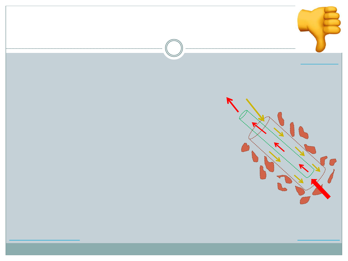
5A. The brain tissue fluid is directly aspirated and the
concentration of the metabolites measured
This is incorrect
The micro-dialysis probe is
essentially a coaxial catheter with
a semipermeable dialysis
membrane lining its tip
Through the outer channel, fluid,
isotonic to the brain extracellular
fluid, is pumped at 0.3µL/min and
aspirated back through the inner
tube
The dialysis membrane at the tip
allows diffusion of water and
solutes from the interstitial fluid
into the catheter along its
concentration gradient
Isotonic fluid
Micro dialysate
Brain tissue
Metabolites &
solutes
Previous Question
Try Again
References
5B. It is used as a test to confirm secondary brain
injury after it is evident on other monitors
Cerebral micro-dialysis can detect
changes in the metabolism at the
cellular level before changes are
detected in other monitors for
brain physiology
The micro-dialysis probe is
essentially a coaxial catheter with a
semipermeable dialysis membrane
lining its tip that allows diffusion
of cellular metabolites
Isotonic fluid
Micro dialysate
Brain tissue
Metabolites &
solutes
Previous Question
Try Again
References

5C. A high Lactate-Pyruvate ratio indicates
cerebral ischemia
Metabolites and solutes measured by cerebral micro-dialysis
Energy related metabolites – glucose, lactate, pyruvate
Markers of secondary brain ischemia
Glucose <1.5 mmol/L
Raised lactate to pyruvate ratio (>20)
Neurotransmitters – glutamate, aspartate
High levels are seen in secondary cerebral ischemia.
Cellular degradation markers – glycerol, potassium
Glycerol is produced by degradation of the phospholipids
from dead cells. High levels have been measured after
severe TBI and also secondary ischemia.
Exogenous – drugs
Previous Question References
Back to Question

5D. A rise in glucose in the micro-dialysate 2-3 days
after a brain injury indicates cell death
This is incorrect.
A rise in glycerol indicates cell death
Glucose level decreases with brain ischemia
Metabolites and solutes measured by cerebral micro-dialysis
Energy related metabolites – glucose, lactate, pyruvate
Markers of secondary brain ischemia
Glucose <1.5 mmol/L
Raised lactate to pyruvate ratio (>20)
Neurotransmitters – glutamate, aspartate
High levels are seen in secondary cerebral ischemia.
Cellular degradation markers – glycerol, potassium
Glycerol is produced by degradation of the phospholipids from dead cells. High levels
have been measured after severe TBI and also secondary ischemia.
Exogenous – drugs
Previous Question
Try Again
References

References
Elwishi M, Dinsmore J. Monitoring the brain.
BJA Education 2019; 19:54-59
Smith M. Neuromonitoring.
Anaesthesia & Intensive Care monitoring 2008; 9; 187-192
Emergency Neurological Life Support (ENLS) course
Cerebral monitoring in the operating room and the intensive care unit.
Journal of Clinical Monitoring and Computing 2005;19: 1–76
(DOI: 10.1007/s10877-005-0712-z)
THANK YOU
Go to Title, Question 1, 2, 3, 4, 5
